Fluorescent probes selectively taken up by tumorous tissue could be used for early disease detection
Published online 28 July 2016
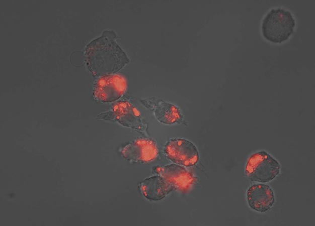
Stimuli-responsive fluorescent polymers light up to reveal the temperature inside cells.
© 2016 Hideko Kanazawa, Keio University
A stimuli-responsive fluorescent polymer that is readily taken up by living cells could become a useful diagnostic tool for identifying diseased cells, Keio University scientists suggest. The research team developed fluorescent polymers whose light output dims in response to warmer temperatures and more acidic pH ― conditions typical of pathological tissue in patients with diseases ranging from cancer to Alzheimer's disease.
The polymers' responsive properties could eventually be used to selectively deliver drugs to diseased cells.
At the heart of the polymer, developed by Hideko Kanazawa and her team at Keio University's Department of Pharmacy, is a molecule called Poly(N-isopropylacrylamide), or PNIPAAm. This polymer is well known for significantly changing its structure in water above a critical temperature.
At low temperatures, a strong interaction between water molecules and the polar nitrogen-rich amido groups in the PNIPAAm polymer means the polymer spreads out into an 'expanded coil' formation. But, warming the solution disrupts the water-polymer interaction, until the critical temperature of 32°C is reached and the polymer suddenly contracts, forming a compact globular structure.
Kanazawa and her team created four new forms of PNIPAAm polymer that incorporate different fluorescent side-chains. Previous work showed these fluorescent groups are pH-responsive, dimming under acidic conditions. But the team showed that, once combined with the PNIPAAm polymer, they became responsive to temperature also. Below the polymer's critical temperature the side chains fluoresce brightly. But as soon as the polymer collapses, the fluorescence is switched off1.
This responsive behavior remains active inside living cells, Kanazawa showed. The researchers attached a non-polar lipid additive called DOPE to the polymer to help draw it through the non-polar cell membrane and into the living cell. They were surprised to find just how effective the strategy proved. "DOPE caused a strong preference for cellular uptake, allowing the polymers to be rapidly internalized without the requirement for a delivery system," Kanazawa says.
The team is already working on new versions of the fluorescent polymer with enhanced functionality, Kanazawa adds. For example, it has developed versions of the polymer that, under the mildly acidic pH around diseased cells, switch from a polar to a non-polar state. This polarity switch dramatically boosts the rate at which the fluorescent probe is taken up by diseased cells compared to healthy ones. "This polymer may offer the potential for development as a diagnosis tool for early tumor detection," Kanazawa says. "Furthermore, it could be used as a tumor-selective intracellular drug delivery system."
Reference
- Yamada, A., Hiruta, Y., Wang, J., Ayano, E. & Kanazawa, K. Design of environmentally responsive fluorescent polymer probes for cellular imaging. Biomacromolecules 16, 2356-2362 (2015). | article