Improved coherent X-ray diffraction imaging makes it possible to analyze the internal parts of cells
Published online 6 March 2015
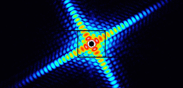
X-ray diffraction pattern from a cube-shaped copper oxide particle, approximately 500 nanometers in size.
© 2015 Masayoshi Nakasako, Keio University
By employing a ring-shaped mask and appropriate signal processing, scientists in Japan have found a way to overcome a common limitation of coherent X-ray diffraction imaging and dramatically improve the quality of analysis1. As a result, they have succeeded in using the imaging technique to analyze the internal structures of biological cells and a collection of metallic nanoparticles.
Masayoshi Nakasako from the Department of Physics at Keio University and the RIKEN SPring-8 Center and his co-workers have been using the X-ray free electron laser at the SACLA facility in Japan for their experiments.
Imaging the diffraction patterns that result when X-rays interact with a sample is a very powerful approach for visualizing the structure of materials. Indeed, the discovery of the double helix structure of DNA was based on analysis of an X-ray diffraction image, the famous 'photo 51' in 1952.
X-rays can deeply penetrate into materials beyond the reach of electron microscopy and their short wavelength yields an imaging resolution of just a few tens of nanometers, far better than optical microscopy. Applying phase retrieval algorithms to the two-dimensional X-ray diffraction pattern captured on an image sensor with an array of pixels allows the electron density map of the sample to be reconstructed.
However, there's a problem that hinders coherent X-ray diffraction imaging experiments that are based on X-ray free electron lasers, especially those experiments involving clusters of metal particles with large scattering cross-sections or cellular organelles. At small diffraction angles, the image sensor receives such an intense X-ray signal that it saturates the detector, resulting in missing data. Nakasako's team solved the problem by employing an annular mask (see image) and dark-field phase-retrieval.
"Through developing a diffraction apparatus called KOTOBUKI-1 and data processing software, we have succeeded in the structure analyses of bacterial cells and organelles in biology and the determination of the size distribution and the structures of synthesized metal nanoparticles from industry," explains Nakasako. "Recently we have succeeded in visualizing the structure of chloroplasts with the dimension of approximately 1 micron at effective resolutions of 70 nanometers, better than the resolution limit of optical microscopy."
The capabilities of this new form of coherent X-ray diffraction imaging are said to be a valuable addition to the other forms of imaging such as fluorescence or electron microscopy, for example. "The complementary use of [coherent X-ray diffraction imaging] together with other imaging techniques will bridge the resolution gap between cellular and structural biology," concludes Nakasako.
Reference
- Kobayashi, A., Sekiguchi, Y., Takayama, Y., Oroguchi, T. & Nakasako, M. Dark-field phase retrieval under the constraint of the Friedel symmetry in coherent X-ray diffraction imaging. Optics Express 22, 27892-27909 (2014). | article